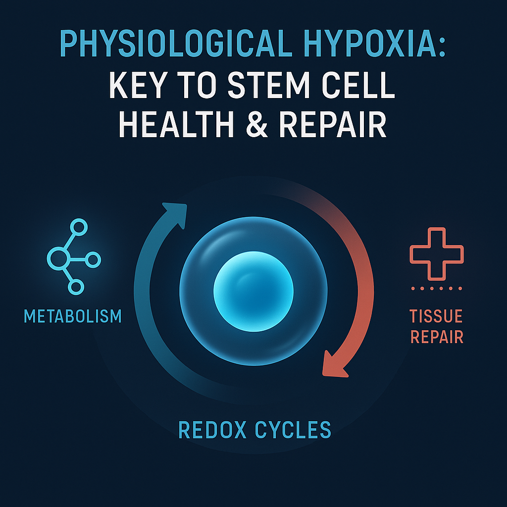
Physiological Hypoxia as a Master Regulator of Stem‐Cell Fate
Physiological hypoxia, characterized by oxygen levels around 3–6% O₂, is crucial for maintaining stem-cell characteristics in various adult regenerative niches [Mas-Bargues et al., 2019]. This low-grade hypoxia is not merely a consequence of suboptimal blood flow; instead, it acts as a vital metabolic signal that preserves stemness across different lineages, including hematopoietic, mesenchymal, neural, and epithelial cells.
HIF Stabilization and Redox Balance
In a low-oxygen environment, hypoxia-inducible factors (HIFs) are stabilized due to inhibited prolyl-hydroxylase activity, allowing for an elevation in the transcription of essential stemness genes such as OCT4, SOX2, KLF4, and NANOG. This process concurrently represses key cell-cycle inhibitors like p16^Ink4a and p21^Cip1, thus delaying the onset of senescence and maintaining genomic integrity [Mas-Bargues et al., 2019]. Additionally, the low oxygen environment induces a variety of antioxidant genes, which help mitigate reactive oxygen species (ROS) accumulation that could otherwise lead to DNA damage.
Metabolic Re-wiring—Glycolysis Over OXPHOS
Hypoxia shifts the energy production pathway from oxidative phosphorylation (OXPHOS) to anaerobic glycolysis, mediated by HIF-1α, which up-regulates pyruvate dehydrogenase kinase (PDK1/4), lactate dehydrogenase A (LDHA), and glucose transporter 1 (GLUT1) [Takubo et al., 2013]. This metabolic shift lowers the flux through the electron transport chain and minimizes ROS leakage. It has been noted that the genetic deletion of PDK1 can lead to the collapse of cellular quiescence in mouse hematopoietic stem cells. Similar enhancements have been documented in human mesenchymal stem cells (MSCs), which show improved clonogenic yield and enhanced tri-lineage differentiation when cultured in 3–5% O₂ [Mas-Bargues et al., 2019].
ROS as Signaling Rheostats
Excessive ROS levels from chronic hyperoxia (>10% O₂) can force lineage commitment; however, controlled “ROS bursts” generated under physiological hypoxia play a critical signaling role. These bursts, activated via mechanisms such as NOX4 or mitochondrial uncoupling, serve as second messengers in critical cellular pathways, including Notch, PI3K-AKT-FOXO, and p38-MAPK [Mas-Bargues et al., 2019]. Consequently, the stem-cell niche operates more like a redox rheostat, allowing for a delicate balance between proliferation and differentiation.
Implications for Ex-vivo Expansion
When cultured at atmospheric O₂ levels (approximately 21%), MSCs, dental pulp stem cells (DPSCs), or neural stem cells (NSCs) face hyperoxic stress. This condition can significantly accelerate telomere erosion and increase rates of chromosomal abnormalities, ultimately halving their colony-forming efficiency [Estrada et al., 2012]. Conversely, continuous culture at 5% O₂ mirrors physiological conditions, restoring glycolytic flux while maintaining mitochondrial membrane potential, facilitating long-term clinical-grade cell preservation with minimal karyotypic drift.
Niche Engineering and Bioreactor Design
Innovative scaffolding solutions should replicate oxygen gradients, with concentrations around 2% O₂ at their core and approximately 6% O₂ at the periphery, closely mimicking the natural bone marrow environment. Furthermore, dynamic bioreactors maintained at 5% O₂ during induced pluripotent stem cell (iPSC) reprogramming have been shown to significantly reduce the latency of reprogramming and allow for the use of dual-factor combination cocktails like OCT4 and KLF4, thus minimizing oncogenic risk [Yoshida et al., 2009].
Hyperbaric Oxygen Therapy: Modulation of Vasculogenic Stem Cells
The application of hyperbaric oxygen (HBO₂) therapy significantly alters the bio-distribution and phenotype of vasculogenic stem/progenitor cells, particularly circulating CD34⁺/VEGFR2⁺ cells [Thom et al., 2006]. These progenitor cells are essential for orchestrating post-natal vasculogenesis, mobilized during treatment in conjunction with a potent pro-regenerative program in wound bed environments.
Oxidative Mobilization in Bone Marrow
A single session of hyperbaric oxygen therapy for 90 minutes elevates arterial O₂ levels to over 2,000 mm Hg, stimulating a regulated ROS/RNS release within the marrow stroma. These oxidative molecules induce a cascade effect, releasing a 2- to 8-fold increase in circulating SPCs within a few hours while avoiding a pro-thrombotic state due to leukocytosis [Thom et al., 2006].
Chemotaxis and Niche Reprogramming at the Wound
At the wound margins, typically characterized by O₂ levels below 10 mm Hg, HBO₂-induced ROS can enhance local concentrations of HIF-1α, VEGF-A, and other key chemokines like SDF-1, facilitating SPC homing and stimulating endothelial differentiation [Fosen & Thom, 2014]. Additionally, the accumulation of lactate from hypoxic glycolysis further stabilizes HIF-1α, creating a feedback loop that amplifies gene expression associated with neovascularization.
Clinical Impact of HBO₂ Therapy
Randomized controlled trials centered on refractory diabetic foot ulcers have demonstrated that adjunctive HBO₂ significantly reduces the time to wound closure and major amputation rates by approximately 50% due to enhanced mobilization of stem cells and improved granulation [Londahl et al., 2010]. In studies of myocardial infarction models, the cyclic application of HBO₂ has led to increased EPC counts, enhanced vascular density, and improved ejection fractions through NOS3-HIF-VEGF pathways [Khan et al., 2009].
Engineering and Translational Trajectories
Emerging strategies include ROS-responsive hydrogels designed to release chemokines like SDF-1 and VEGF in synchronization with hyperbaric oxygen therapy, enhancing the recruitment of stem cells and promoting neovascularization [Londahl et al., 2010]. Additionally, pre-conditioning autologous MSC/EPC grafts with pre-therapy hypoxia prior to HBO₂ has shown to significantly improve engraftment in ischemic tissues.
Oxygen-Sensitive Checkpoints in Stem Cells
Oxygen-regulated checkpoints play pivotal roles in stem cell fate decisions. Prolyl-hydroxylases and factor-inhibiting HIF (FIH) act as molecular gatekeepers that can modulate HIF activity based on ambient pO₂ [Mas-Bargues et al., 2019]. At pO₂ levels above 5-7%, hydroxylation of HIF-α isoforms leads to their degradation. This allows HIF-1/2α to accumulate and activate transcriptional programs crucial for maintaining stem cell quiescence under suboptimal oxygen conditions.
Metabolic Checkpoints—PDK, mTOR, and FOXO
HIF-induced metabolic regulation occurs through pathways involving PDK1/4, which inhibit pyruvate dehydrogenase activity, forcing cells towards glycolysis instead of oxidative metabolism. This is critical for maintaining stem cells in a replenishing state [Takubo et al., 2013]. Moreover, under hypoxic conditions, mTORC1 activity is suppressed, leading to preserved autophagy and enhancing cellular longevity and genomic stability.
Redox Pulses as Signaling Nodes
Sub-micromolar ROS pulses act as important regulatory signals, enabling the fine-tuning of cellular responses through pathways such as PI3K-AKT-FOXO and p38-MAPK. However, excessive ROS exposure can lead to oxidative stress, which may permanently commit cells to senescence or apoptosis, thus underscoring the delicate balance required in therapeutic interventions [Takubo et al., 2013].
SDF-1/CXCR4 Axis—Linking Systemic Mobilization to Local Homing
HIF-1α plays an essential role in upregulating the SDF-1 chemokine during hypoxic conditions, while HBO₂-induced ROS enhances CXCR4 expression on circulating SPCs. This results in the precise homing of stem cells toward hypoxic regions, facilitating tissue repair while preventing premature differentiation until necessary [Milovanova et al., 2009].
Translational Leverage Points
Innovative strategies, such as transient PHD (prolyl hydroxylase domain) inhibitors, can amplify endogenous HIF-Notch signaling. This approach can expand stem-cell pools pre-operatively or prior to chemotherapy, potentially enhancing outcomes in regenerative therapies.
Engineering Stem-Cell Therapeutics
Scaffold design and composition must account for oxygen architecture, incorporating β-tricalcium-phosphate scaffolding alongside human dental pulp stem cells, significantly improving osteogenic activity under controlled hypoxia [Vina-Almunia et al., 2017]. Additionally, preconditioning regimens can up-regulate survival factors in MSCs, doubling their capacity for bone formation in critical defects [Volkmer et al., 2010].
Future Prospects and Corridors of Innovation
The advent of ROS-responsive hydrogels and wearable pO₂ sensors represents a paradigm shift in regenerative medicine, enabling precise control over stem-cell activity and therapeutic efficacy. By bridging gaps between oxygen dynamics and stem-cell functionality, next-generation tools are set to refine strategies that maximize tissue repair and regeneration across a range of clinical applications.
