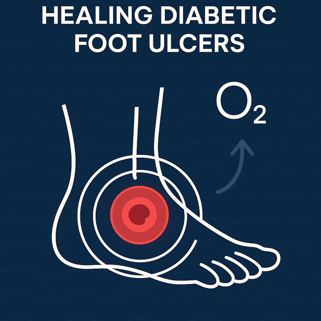
Pathophysiology of the Diabetic Wound & the HBOT Rationale
Diabetic foot ulcers (DFUs) represent a significant challenge due to their multifaceted pathophysiology. Chronic hypoxia contributes to this issue when conditions such as peripheral arterial disease and capillary basement-membrane thickening cause transcutaneous oxygen tensions (TcPO₂) to fall below the critical 30 mmHg threshold necessary for fibroblast proliferation and collagen cross-linking. Goldman’s systematic review highlights that over 70% of relapsing lesions exhibit critically low TcPO₂ levels, which can only be normalized through hyperbaric oxygen therapy (HBOT), rather than through normobaric oxygen supplementation [Goldman 2009].
Furthermore, sustained inflammation and oxidative stress, manifested through persistent macrophage activation under high glucose conditions, lead to rampant TNF-α and IL-1β production. A study by Capó et al. illustrated that 20 sessions of HBOT significantly reduced these inflammatory markers and improved the overall antioxidant capacity in diabetic wound plasma [Capó 2023].
Matrix and angiogenic failures contribute to delayed healing, where matrix-metalloproteinase-9 degradation undermines collagen integrity and endothelial senescence halts angiogenesis. HBOT alleviates these dysfunctions by stabilizing crucial signaling pathways and promoting neovascularization while suppressing MMP-9 activity [Tejada 2019]. This results in a marked increase in collagen deposition in hyperglycaemic skin, as demonstrated by André-Lévigne et al., who noted a significant rise in collagen type I/III ratios [André-Lévigne 2016].
Molecular-Cellular Mechanisms Triggered by HBOT
HBOT initiates a series of molecular and cellular responses crucial for wound healing. The rapid increase in pressure (2.0–2.8 ATA) elevates plasma oxygen levels beyond 1,400 mmHg, creating a controlled oxidative environment conducive to healing. This spike enhances redox regulation, activating the Nrf2 pathway, which is essential for increasing various antioxidant enzymes while effectively reducing oxidative damage markers [Goldman 2009]. Capó et al. reiterated that over a 20-session span, HBOT fosters an antioxidant milieu, countering inflammation effectively [Capó 2023].
In inflammatory states characterized by M1 macrophage dominance, HBOT reprograms the immune landscape. By inhibiting NF-κB signaling, HBOT facilitates a transition towards the M2 phenotype, promoting efficient tissue healing through enhanced efferocytosis and improved neutrophil bactericidal activity [Capó 2023]. This supports the documented clinical observations of faster infection clearance post-HBOT [Chen 2017].
Additionally, HBOT augments angiogenic signaling by raising factors such as HIF-1α and VEGF, leading to increased capillary density in diabetic tissues and thus promoting effective healing [André-Lévigne 2016]. Furthermore, it aids collagen metabolism by enhancing the hydroxylation of proline and lysine, which is indispensable for correct collagen structure and tensile strength [van Neck 2017].
Pre-clinical Evidence
Pre-clinical models have offered critical insight into the response of chronic diabetic wounds to HBOT. Van Neck et al. demonstrated in a rat model that those with higher baseline peri-wound TcPO₂ showed much greater re-epithelialization with HBOT treatment, reinforcing the need for physiological baseline evaluation [van Neck 2017]. Conversely, conditions with critically low TcPO₂ yielded minimal improvement from HBOT, showcasing the importance of patient stratification in clinical trials.
Utilizing an ischemic–hyperglycaemic flap model, André-Lévigne et al. noted significant increases in collagen type I/III deposition and marked improvements in tissue perfusion from HBOT, although some subjects displayed a lack of response due to existing endothelial dysfunction [André-Lévigne 2016].
Moreover, in larger animal models, Tejada’s review highlights that while some exhibited strong angiogenic responses to HBOT, others presented limited improvements due to advanced microvascular complications [Tejada 2019]. These findings emphasize the necessity of identifying potential responders pre-HBOT.
Clinical-Trial & Meta-analytic Signal Strength
| Study | Design / n | HBOT Protocol | Primary Endpoint | Key Result |
|---|---|---|---|---|
| Goldman et al. 2009 | Systematic review (8 trials, 447 DFUs) | 2.0–2.8 ATA, 30–40 dives | Limb salvage & wound closure | Pooled odds of major amputation fell by 40 %; time-to-healing shortened by 2–4 weeks [Goldman 2009] |
| Löndahl et al. 2011 | Double-blind RCT / 75 | 2.5 ATA, 90 min, 40 dives | Complete ulcer closure at 1 yr | 61 % HBOT vs 27 % sham (p = 0.009); SF-36 physical score +8.4 ± 3.7 points [Löndahl 2011] |
| Chen et al. 2017 | Open-label RCT / 94 | 2.5 ATA, 80 min, 40 dives | Closure at 12 wks | 52 % HBOT vs 29 % control (RR = 1.8, p = 0.03) [Chen 2017] |
| Lam et al. 2017 | Meta-analysis (9 RCTs) | — | Major amputation | Pooled risk ratio 0.58 (95 % CI 0.36–0.92) [Lam 2017] |
The clinical evidence consistently supports HBOT’s efficacy, revealing higher healing rates and notable reductions in amputation rates when protocols achieve sufficient peri-wound TcPO₂ levels [Capó 2023].
Practical Protocol Optimisation
To maximize HBOT’s efficacy for DFU management, specific session parameters should be adhered to. The evidence-backed ranges for HBOT include pressures between 2.0–2.8 ATA, primarily noted at 2.4–2.5 ATA. Keeping oxygen concentration at 100% via masks or hoods ensures maximal diffusion into the tissues. Sessions should last between 60–90 min to follow established regimens found effective in clinical trials [Chen 2017].
Frequent sessions, ideally five per week, maintain pro-angiogenic signaling, while a course length of at least 30 dives is recommended for optimal healing [Lam 2017].
Physiological assessments such as baseline TcPO₂ testing before the treatment can be crucial. Wounds displaying TcPO₂ above 25 mmHg have yielded optimal healing responses, offering a strategy for predicting treatment outcome [Goldman 2009]. Adapting treatment protocols, including escalating pressure for non-responders, can further enhance clinical outcomes.
Safety & Contra-indications
The safety profile of HBOT is largely favorable, with adverse events mostly mild and resolving spontaneously. Common occurrences include middle-ear barotrauma at rates of 1–3%, temporary myopia in ≤5%, and a very low incidence of oxygen-induced seizures at about 0.01% [Goldman 2009] [Chen 2017].
Contraindications focus primarily on specific conditions such as untreated pneumothorax and concurrent chemotherapy with certain agents due to the risk of pulmonary complications. Safety protocols and regular assessments before and during treatment sessions are vital to mitigate risks [Capó 2023].
Frontier Research Directions
The evolving landscape of HBOT adapts to emerging insights, emphasizing precision stratification through multi-omic profiling. This includes using pre-treatment TcPO₂ alongside transcriptomic panels to improve predictive outcomes for HBOT responsiveness [van Neck 2017]. Additionally, exploring combination therapies, such as integrating HBOT with growth factor applications or stem cell therapies, may enhance the regenerative capacity of diabetic wounds [Tejada 2019].
Furthermore, the integration of innovative monitoring systems such as wearable TcPO₂ sensors can help tailor treatment by offering real-time metrics on oxygen dynamics. Machine-learning models based on these data might forecast healing probabilities more accurately, leading to more personalized treatment pathways [Capó 2023].
Key Take-home Messages
HBOT emerges as a transformative adjunct in managing diabetic foot ulcers, displaying a well-supported mechanistic foundation and strong clinical efficacy evidenced by numerous rigorous studies. Optimized HBOT protocols — utilizing appropriate pressures and session frequencies — can mitigate DFU complexities through enhanced angiogenesis, reduced inflammation, and improved collagen synthesis [Goldman 2009]. The ability to personalize treatment regimens while maintaining focus on the patient’s baseline physiological status will be instrumental in enhancing therapeutic outcomes in future trials [Chen 2017].
Sources
- Bentham Direct – Therapeutic effects of hyperbaric oxygen in the process of wound healing
- MDPI – Hyperbaric oxygen therapy reduces oxidative stress and inflammation, and increases growth factors favouring the healing process of diabetic wounds
- LWW – Adjunctive hyperbaric oxygen therapy for healing of chronic diabetic foot ulcers: a randomized controlled trial
- Wiley Online Library – Hyperbaric oxygen therapy promotes wound repair in ischemic and hyperglycemic conditions, increasing tissue perfusion and collagen deposition
- ScienceDirect – Hyperbaric oxygen therapy for wound healing and limb salvage: a systematic review
- PLOS ONE – Hyperbaric oxygen therapy for wound healing in diabetic rats: Varying efficacy after a clinically-based protocol
- Wiley Online Library – Hyperbaric oxygen therapy improves health‐related quality of life in patients with diabetes and chronic foot ulcer
- ASWC Journal – Hyperbaric oxygen therapy: exploring the clinical evidence
- Wiley Online Library – Hyperbaric oxygen therapy improves health‐related quality of life in patients with diabetes and chronic foot ulcer
- ASWC Journal – Hyperbaric oxygen therapy: exploring the clinical evidence
